

Our Technology
How We Can Help
Here at Kapperman McGarvey Eye Group, we use cutting-edge technology to assist in diagnosing any eye conditions that may affect your vision or the health of your eyes.

Technology That Works for You

Dry Eye Solution with Lumenis Optilight IPL
Millions of people suffer from eye and vision-related problems. For some, the solution is as simple as wearing prescription eyeglasses or contact lenses. However, others face challenges that aren’t as easy to rectify. That’s where Lumenis comes into play.

Optical Coherence Tomography (OCT)
The OCT uses near infrared light to provide detailed images of the retinal layers. This device helps to identify:
Retinal holes and tears
Macular degeneration
Macular edema
Glaucoma

Humphrey Visual Field Device (HVF)
The HVF measures the extent of a patient's field of vision.This device is used for detection of defects in the visual field related to:
Glaucoma
Brain injury
Pathology in the brain
Droopy eyelids
Multiple Sclerosis or damage to the optic nerve
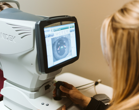
NIDEK
The NIDEK is an advanced vision-assessment system that combines topography, wavefront, autorefraction, keratometry, and pupillometry, which allows accurate and reliable analysis of corneal aberration. This device helps by:
Showing a point-by-point map of eye aberrations.
Gives many more data points than other aberrometers — allowing precise mapping of irregular astigmatisms.
Improving the accessibility to the eye.

Digital Retinal Imaging (DRI) with Optomap
At Kapperman McGarvey Eye Group, we believe that one of the most important components of the eye exam is retinal imaging. Like dentists use X-rays to evaluate the underlying health of teeth, optometrists use DRI to evaluate the underlying health of the eye. Not only does DRI provide your doctor with a better view of the retina, it gives them a permanent record of that view. Baseline imaging is invaluable when it comes to identifying whether a problem is longstanding or advancing. We recommend DRI with Optomap every time you get an eye exam. In some instances, the doctor may deem it necessary to dilate your eyes even if you have done DRI. In every situation, the doctor will make this decision based on what is best for the health of your eyes.
Optical Coherence Tomography (OCT)
The OCT uses near infrared light to provide detailed images of the retinal layers. This device helps to identify:
Retinal holes and tears
Macular degeneration
Macular edema
Glaucoma
Humphrey Visual Field Device (HVF)
The HVF measures the extent of a patient's field of vision.This device is used for detection of defects in the visual field related to:
Glaucoma
Brain injury
Pathology in the brain
Droopy eyelids
Multiple Sclerosis or damage to the optic nerve
Ready for Cutting-Edge Eye Care?
We use the latest technology to evaluate your eyes so nothing goes by unnoticed.

See what eye conditions we can treat

Get Comprehensive & Dedicated Eyecare


Keep
In Touch
Contact Info
Phone:
(423) 892-2020
(423) 892-2020
Fax: (423) 855-0329
Hours of Operation
- Monday 7:30am - 5:30pm
- Tuesday 7:30am - 5:30pm
- Wednesday 7:30am - 5:30pm
- Thursday 9:00am - 5:30pm
- Friday 7:30am - 5:30pm
- Saturday Closed
- Sunday Closed
© 2026 Kapperman McGarvey Eye Group. All rights Reserved. Accessibility Statement - Privacy Policy - Sitemap
Powered by: